A dental granuloma is one of the more common findings on a routine dental X-ray — yet most patients have no idea it is there. It forms silently at the root of a tooth, triggered by a chronic bacterial infection, and grows slowly over months or even years without producing obvious pain. By the time it does cause symptoms, the infection has often been spreading unchecked for a significant period.
Understanding what a dental granuloma is, what causes it, and how it is treated can help you seek care early and protect your teeth from avoidable complications.
What Is a Dental Granuloma?
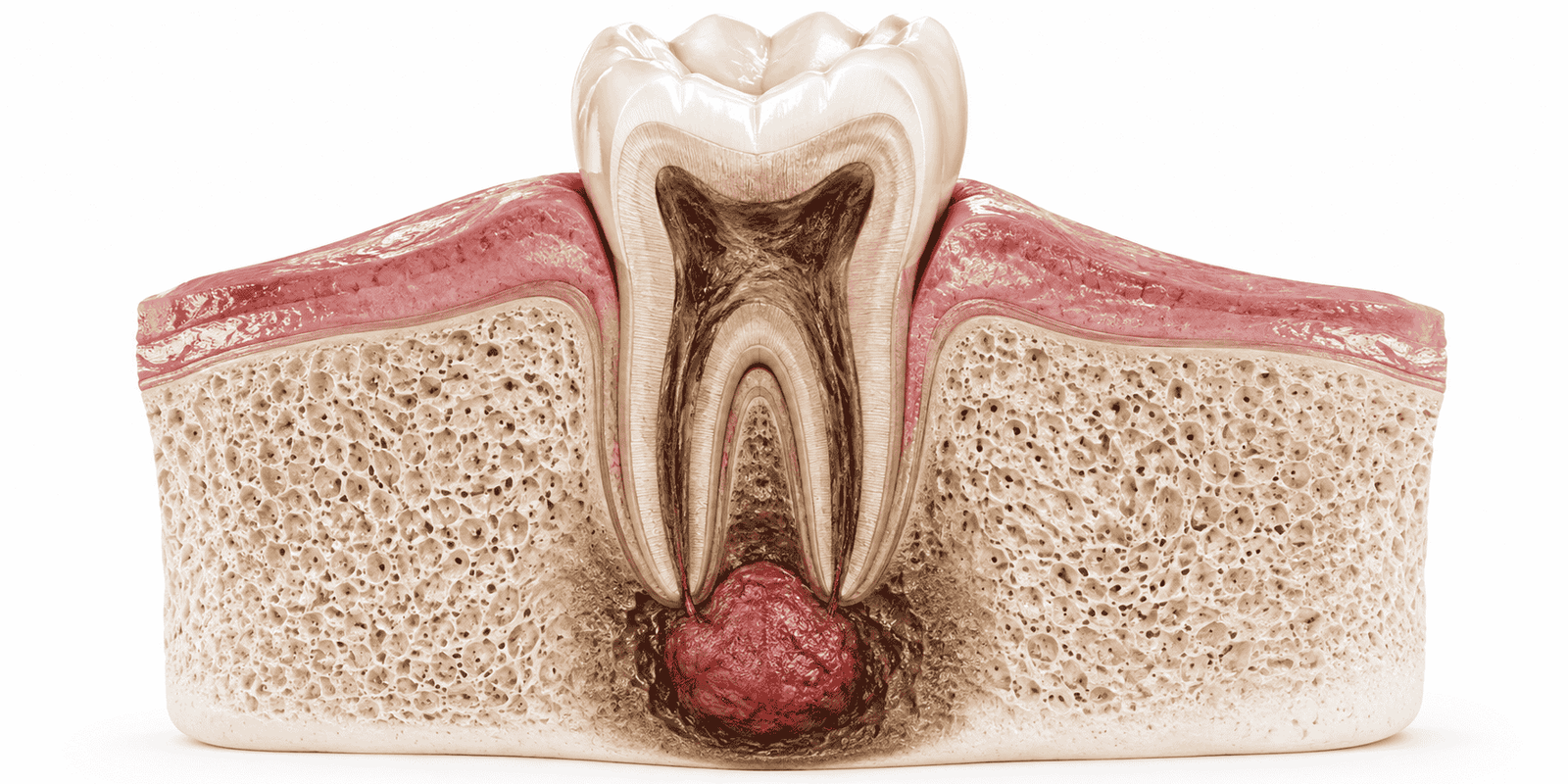
A dental granuloma is a localized mass of chronically inflamed tissue that forms at the apex of a tooth root. It develops when the immune system responds to a persistent bacterial infection inside the tooth pulp by building a wall of immune cells around the infected site. This walled-off mass is the granuloma. Rather than eliminating the infection, it contains it — at the cost of ongoing tissue damage and slow bone loss around the root tip.
Periapical Granuloma vs. Dental Cyst
A periapical granuloma and a dental cyst are related conditions that can appear similar on an X-ray, but they are structurally different and respond differently to treatment.
A granuloma is a solid mass of inflamed granulation tissue that forms at the root tip. It is primarily managed through root canal treatment, which eliminates the bacteria driving the immune response.
A cyst is a fluid-filled sac with an epithelial lining. It develops when a long-standing granuloma undergoes cystic transformation. Dental cysts are more likely to require surgical intervention because root canal treatment alone may not fully resolve them.
Dental Granuloma vs. Abscess
A granuloma and an abscess both originate from infected dental pulp, but they differ in how they develop and how urgently they require treatment.
A dental granuloma develops slowly over weeks or months. The body’s immune system partially controls the infection by encapsulating it, which is why the condition often produces little or no pain in its early stages. However, the infection remains active underneath, continuing to destroy surrounding bone tissue.
An abscess is an acute, rapidly developing infection. It produces a pocket of pus, severe pain, facial swelling, and sometimes fever. An abscess demands urgent dental treatment because the infection can spread to the jaw, neck, and beyond. A granuloma that is left untreated can break down and convert into an acute abscess.
What Causes a Dental Granuloma?
Several clinical pathways lead to granuloma formation at the root tip. In all cases, the underlying mechanism is the same: bacteria reach the periapical tissues through the root canal system, and the immune system responds by forming granulation tissue around the infected site.
1. Untreated Tooth Decay
Advanced tooth decay that is not treated in time allows bacteria to penetrate the tooth pulp. Once inside the pulp chamber, they move down the root canals toward the apex. The pulp tissue gradually dies, removing the tooth’s natural ability to fight infection, and the bacteria begin colonising the bone at the root tip.
Many patients notice the toothache that accompanied active pulp infection suddenly disappears — this is not recovery.
It often means the pulp has become necrotic, and the infection has progressed beyond the tooth into the surrounding bone. A tooth filling placed at the first sign of decay prevents the cavity from ever reaching this stage.
2. Incomplete or Failed Root Canal Treatment
Root canal treatment that does not fully clean and seal all canals can leave residual bacteria inside the root system. These bacteria continue to leach out through the root apex and sustain a low-grade inflammatory response in the periapical bone.
Over time, this results in granuloma formation at the treated tooth. Retreatment or, in resistant cases, a surgical procedure called apicoectomy is required to resolve the infection.
3. Dental Trauma
A significant blow to a tooth — from a fall, a sports injury, or any impact — can disrupt the blood supply to the pulp without visibly fracturing the tooth. The pulp may die months or years later as a delayed consequence of the trauma. Once the pulp is non-vital and the canal becomes contaminated, the conditions for granuloma formation at the root apex are established.
4. Persistent Bacterial Infection
Chronic bacterial infection at the root apex, whether from decay, failed treatment, or trauma, sustains granuloma growth over time. Rather than resolving, the granuloma continues to enlarge, progressively destroying the surrounding alveolar bone. If the immune response becomes overwhelmed, the chronic inflammation can convert into an acute exacerbation, presenting as a painful swelling or abscess with or without a draining sinus tract on the gum.
Signs and Symptoms of Dental Granuloma
Dental granuloma symptoms vary considerably between patients depending on the size of the lesion, the health of the surrounding tissues, and how long the condition has been present. When symptoms do appear, they can include persistent dull or throbbing pain in the affected tooth, tenderness when biting or pressing on the tooth, swelling or a slight puffiness in the gum near the root tip, a small pimple-like bump on the gum that may drain intermittently, tooth darkening relative to adjacent teeth, and in more advanced cases, general malaise or a low-grade feeling of unwellness.
Why Some Granulomas Are Asymptomatic
A granuloma remains asymptomatic when the immune system successfully contains the infection within the inflamed tissue mass. As long as the balance holds between bacterial activity and immune response, the surrounding bone and gum tissues continue functioning without producing pain signals.
This balance is fragile. A systemic illness, immunosuppression, or a period of physical stress can tip the balance, causing a previously silent granuloma to become acutely inflamed and painful. This is why dentists emphasise regular X-ray monitoring even when a tooth feels completely fine.
Price Guides
- Hollywood Smile Price
- Metallic Braces Price
- Porcelain Veneers Price
- Wisdom Tooth Extraction Price
- Root Canal Price
Smile Makeover Cost - Teeth Cleaning Price
- Teeth Cap Price
- Dental Bridge Price
- Zirconia Crown Price
How Is a Dental Granuloma Diagnosed?
Diagnosis begins with a clinical examination, but a clinical examination alone is rarely sufficient to confirm a granuloma. Your dentist will need imaging to visualise the periapical area clearly and determine the size and extent of the lesion.
Dental X-Rays
A periapical tooth X-ray is the standard first-line investigation for a suspected dental granuloma. On the X-ray, the granuloma appears as a well-defined, rounded dark area at the root tip. This dark area represents the low-density inflamed tissue and bone resorption surrounding the apex.
The size, shape, and sharpness of the lesion’s borders help the dentist assess whether it is more likely to be a granuloma, a cyst, or an abscess.
CBCT Scan for Complex Cases
A cone beam computed tomography scan provides a three-dimensional view of the periapical area and is particularly useful when the granuloma is in a location where standard X-rays are limited, such as posterior roots, multi-rooted teeth, or when the lesion is adjacent to important anatomical structures like the maxillary sinus or the inferior dental nerve.
CBCT scanning allows precise assessment of bone loss extent and helps plan surgical treatment when it is required — including procedures like sinus lifting or membrane grafting when bone reconstruction is needed after the granuloma is resolved.
Clinical Examination
The clinical examination includes percussion testing of the tooth to check for tenderness, palpation of the gum over the root apex, and assessment of any sinus tract present on the gum surface. Cold and electric pulp testing determine whether the tooth pulp is still vital.
A tooth with a periapical granuloma is typically non-responsive to vitality testing, confirming that the pulp is no longer alive and that the infection is coming from a necrotic canal system.
4 Treatment Options for Dental Granuloma
Treatment selection depends on the size of the granuloma, the restorability of the tooth, and the presence of any complications such as a cyst, abscess, or significant bone loss.
1. Root Canal Treatment
Root canal treatment is the first-line treatment for most dental granulomas. The dentist accesses the root canal system, removes all infected and necrotic pulp tissue, disinfects the canals with irrigating solutions, shapes the canal walls, and seals the canals with a biocompatible filling material.
2. Apicoectomy
When root canal treatment has already been performed and the periapical lesion persists, or when the root canal cannot be accessed or retreated effectively, an apicoectomy is performed.
This is a minor oral surgical procedure in which the dentist makes a small incision in the gum, reflects the tissue to expose the root apex, removes the infected apical portion of the root along with the surrounding granulomatous tissue, and places a root-end filling to seal the apex.
3. Tooth Extraction
When the tooth is extensively damaged, has poor bone support, or has failed previous attempts at root canal treatment and apicoectomy, tooth extraction becomes necessary. Retaining a tooth with a large, unresolvable granuloma carries ongoing risk of infection spread to adjacent teeth and bone.
Once the tooth is extracted and the socket is thoroughly curetted, patients can explore tooth replacement options including dental implants, crowns and bridges, or dentures.
4. Antibiotic Support
Antibiotics are not a standalone treatment for dental granuloma. They cannot penetrate the granulomatous tissue in concentrations sufficient to eliminate the bacteria driving the infection.
However, antibiotics are prescribed in specific circumstances — when there is an associated acute abscess with spreading infection, significant facial swelling, or signs of systemic involvement such as fever. They are always used alongside definitive dental treatment, never as a substitute for it.
What Happens If a Dental Granuloma Is Left Untreated?
A dental granuloma does not resolve on its own. Without treatment, it continues to grow and cause progressive damage.
An untreated granuloma can gradually destroy the bone around the root tip. In some cases, it may develop into a dental cyst, which can be harder to treat with root canal therapy alone.
The infection can also lead to an acute abscess, causing pain, swelling, and potentially spreading infection. Continued bone and tissue damage may eventually result in tooth loss.
In severe or neglected cases, the infection can spread into the jawbone and cause osteomyelitis, a serious bone infection.
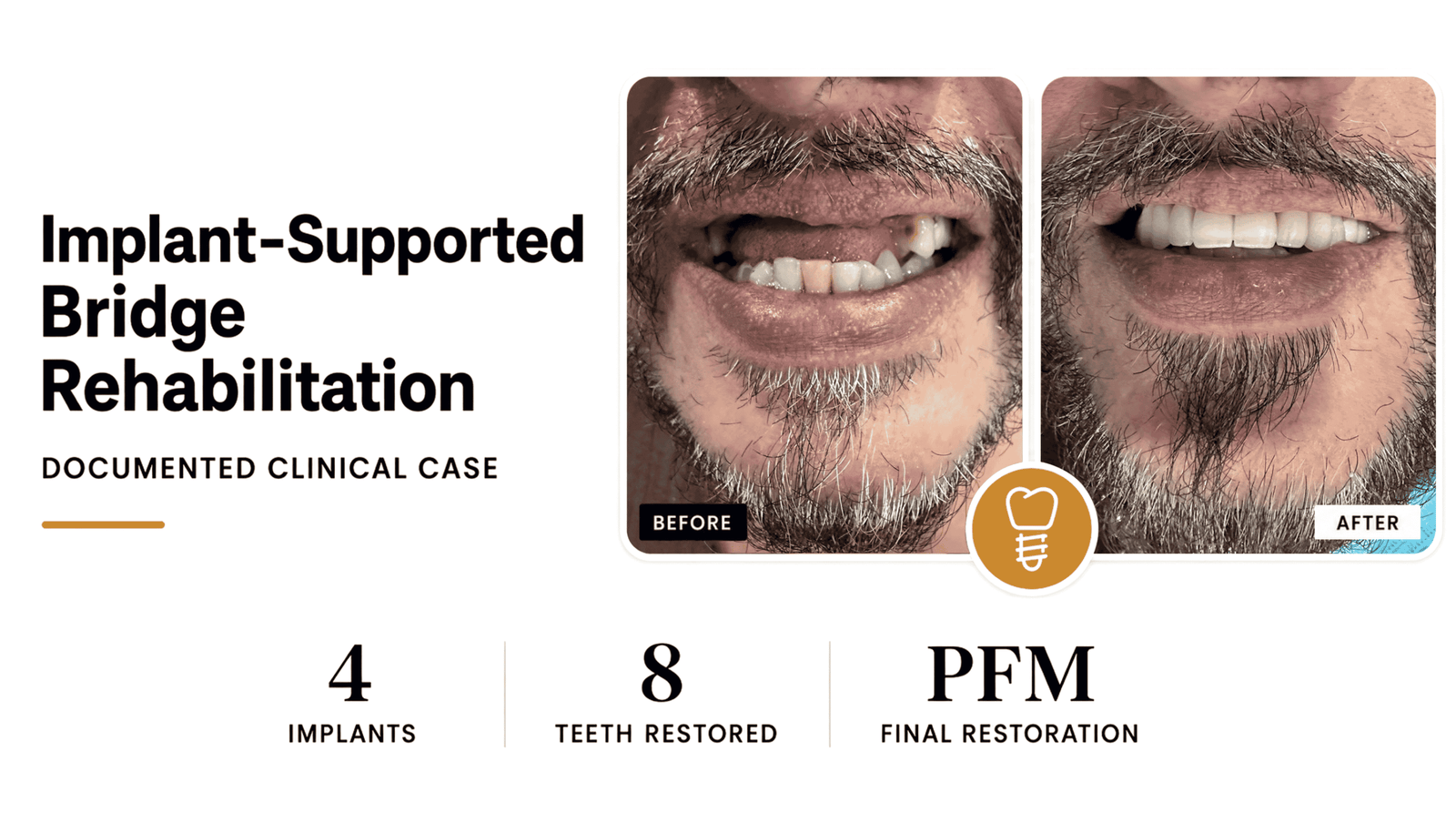
When a tooth cannot be saved and must be extracted, patients are left needing a replacement. Options at that point include dental implants — which are the gold standard for replacing a single missing tooth — or crowns, bridges, and dentures. Patients requiring implant placement after significant bone loss from a granuloma often need additional procedures such as bone grafting with membrane grafts or sinus lifting before an implant can be placed.
There is also emerging evidence of links between chronic periapical infection and systemic conditions. Bacteria from untreated periapical lesions can enter the bloodstream intermittently, contributing to low-grade systemic inflammation. For patients in Karachi with existing health conditions, this makes timely treatment even more important.
Services We offer:
Titanium Implants | Swiss Implants | German Implants | Korean Implants
Composite Bonding | Fluoride Treatment | Teeth Gap Closure | Invisible Aligners
Frequently Asked Questions About Dental Granuloma
Is a dental granuloma dangerous?
Yes, particularly if left untreated. While the body’s immune response initially contains the infection, the granuloma does not eliminate it. Over time, the lesion grows, destroys surrounding bone, and can convert into a cyst, an abscess, or a source of spreading infection. Early root canal treatment or surgical management prevents these complications entirely.
Can a dental granuloma heal on its own?
No. A dental granuloma cannot resolve without professional intervention. The bacteria inside the root canal are the source of the problem, and they cannot be removed by the immune system or by antibiotics alone. Only root canal treatment or tooth extraction eliminates the bacterial source and allows the periapical tissues to heal.
Does a dental granuloma always cause pain?
No. Many granulomas are completely painless, particularly in the early and mid stages of development. Pain tends to appear when the lesion becomes large enough to compress adjacent structures, or when the chronic infection flares into an acute phase. This is why regular dental X-rays matter even when your teeth feel fine.
Can a dental granuloma lead to tooth loss?
Yes. Untreated granulomas progressively destroy the bone surrounding the root. Once enough supporting bone is lost, the tooth becomes mobile and extraction becomes the only option. Patients who lose a tooth this way can later restore the gap with a dental implant, a fixed bridge, or a denture depending on their bone condition.
Does treatment for dental granuloma always involve root canal treatment?
Root canal treatment is the most common treatment approach, but not the only one. If the tooth has already been root-treated and the lesion persists, an apicoectomy performed as part of oral surgery is the next step. If the tooth is non-restorable, extraction is necessary. The right approach depends on the size of the lesion and the overall condition of the tooth.
Is treatment for dental granuloma painful?
No. Both root canal treatment and apicoectomy are performed under local anaesthesia, so you should not feel pain during the procedure. Mild discomfort and soreness in the days following treatment are normal and manageable with standard over-the-counter pain relief. Most patients return to normal activities within a day or two.
How long does recovery take after granuloma treatment?
Soft tissue recovery after root canal treatment is relatively quick, usually within a week. Radiographic bone healing at the apex takes three to twelve months. After an apicoectomy, gum healing takes two to four weeks, with complete bone regeneration continuing for several months beyond that.
Can the granuloma come back after treatment?
Yes, recurrence is possible if the root canal was not fully cleaned and sealed, if the tooth sustains new leakage from a failing crown or filling, or if a cyst had formed within the lesion before treatment. This is why follow-up X-rays are essential after treatment, and why thorough initial treatment matters.
When should someone in Karachi see a dentist about a possible granuloma?
If you have a tooth that was previously painful and then stopped hurting for no apparent reason, a tooth that has darkened, any tenderness when biting, or a small recurring pimple on the gum near a tooth, see a dentist promptly. Many granulomas in Karachi are only discovered after they have already caused significant bone loss because patients delay seeking care when the pain subsides. A dental X-ray can identify a granuloma at an early, easily treatable stage.
Get Expert Dental Granuloma Treatment at The Dental Clinic, Karachi
Dental granuloma treatment requires accurate diagnosis, thorough root canal therapy or surgical management, and disciplined follow-up to confirm that the periapical lesion is genuinely healing. At The Dental Clinic in Gulshan-e-Iqbal, patients across Karachi receive the full diagnostic and treatment pathway under one roof, from periapical X-rays and CBCT scanning through root canal treatment and oral surgery, with post-treatment monitoring at every stage.

For teeth that cannot be saved, the clinic’s dental implant and crowns and bridges services handle full restoration in the same practice, along with membrane grafting and sinus lifting for cases that require bone reconstruction before implant placement.
Dr. Saqib Minhas has been in clinical practice since 2009, with postgraduate training and over a decade of experience managing complex endodontic and periapical cases including persistent granulomas, failed root canal retreatments, and teeth that require surgical intervention to save. His approach is thorough and unhurried: every case is assessed individually, treatment options are explained clearly, and no procedure is recommended unless it is clinically justified.